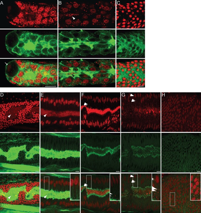
Fig. 7. Two modes of population in the syncytial ovary.
Longitudinal confoncal sections of B. malayi and L. sigmondontis adult ovaries stained with propidium iodide -red- and phalloidin -green-. (A–C) B. malayi female ovary. (A) distal tip, the arrow points to the DTC, (B) more proximal ovary, the arrowheads point to the Wolbachia in the rachis, (C) cellularization zone. (D–H) L. sigmondontis ovary, from distal to proximal ends. Arrowheads point to the Wolbachia. (D–G) syncytial ovary, (H) cellularization. Scale bar = 5 µm.

