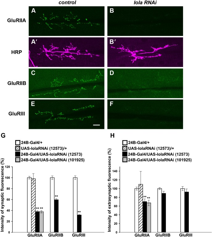
Fig. 3. Synaptic GluR abundance is decreased at the larval NMJ in lola RNAi mutants.
(A–F) Confocal images of NMJs stained for GluRIIA (A,B), GluRIIB (C,D), GluRIII (E,F) and HRP (A′,B′) in control (A,A′,C,E) or in lola RNAi mutants (B,B′,D,F). Scale bar = 20 µm. (G) Quantification of fluorescence intensity for GluRIIA, GluRIIB and GluRIII staining. (H) Extrasynaptic GluR levels in lola RNAi mutants. Data represent % of control fluorescence intensity. **p<0.0005.

