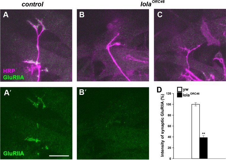
Fig. 4. Synaptic GluRIIA abundance is decreased at the embryonic NMJs in lola mutants.
(A–C) Confocal images of NMJs in dorsal muscles (20 hr AEL) stained for GluRIIA (green) and HRP (magenta) in control (A,A′) and lolaORC46 (B,B′,C) embryos. Scale bar = 10 µm. (D) Quantification of the GluRIIA intensity within the HRP-positive NMJs. **p<0.0005.

