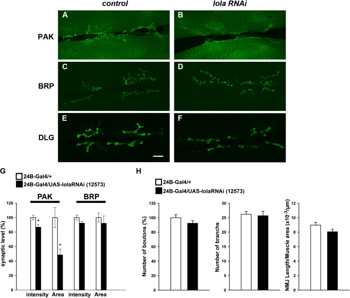
Fig. 6. Postsynaptic PAK and DLG abundance, but not presynaptic BRP abundance, are altered in lola RNAi mutants.
(A–F) Confocal images of NMJs stained for PAK (A,B) and BRP (C,D) and DLG (E,F) in control (A,C,E) and in lola RNAi mutants (B,D,F). Scale bar = 20 µm. (G) Quantification of the fluorescence intensity of PAK and BRP staining (*p<0.05). Data represent % of average fluorescence intensity among control animals. (H) Quantitative analysis of presynaptic morphology in lola RNAi mutants. The number of boutons represent % of the control value, which was 110.6±4.7.

