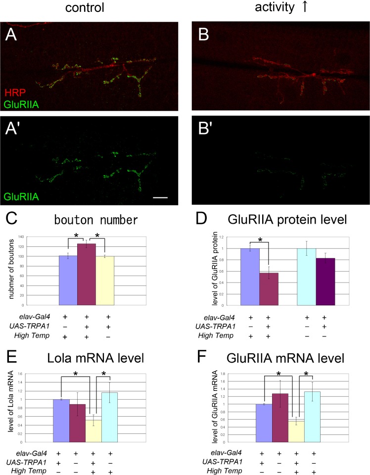
Fig. 9. The transcript levels of lola and GluRIIA decreased after increased activity.
(A–B′) Confocal images of NMJs stained for HRP (A,B) and GluRIIA (A–B′) in control (A,A′) or in a larva stimulated with dTRPA (B,B′). Scale bar = 10 μm. (C,D) Quantification of the number of boutons and relative fluorescence intensity for GluRIIA staining. The number of boutons was increased and the level of GluRIIA was decreased upon dTRPA stimulation with pulses of high temperature shift (High Temp, *p<0.05). The temperature shift itself had no significant effect on these values in the control animals. (E,F) Real-time quantitative PCR analysis of the level of lola (E) and GluRIIA (F) mRNA. The transcript level of Lola and GluRIIA were significantly reduced by increased activity (*p<0.05). Data represent % of control values in C-F.

