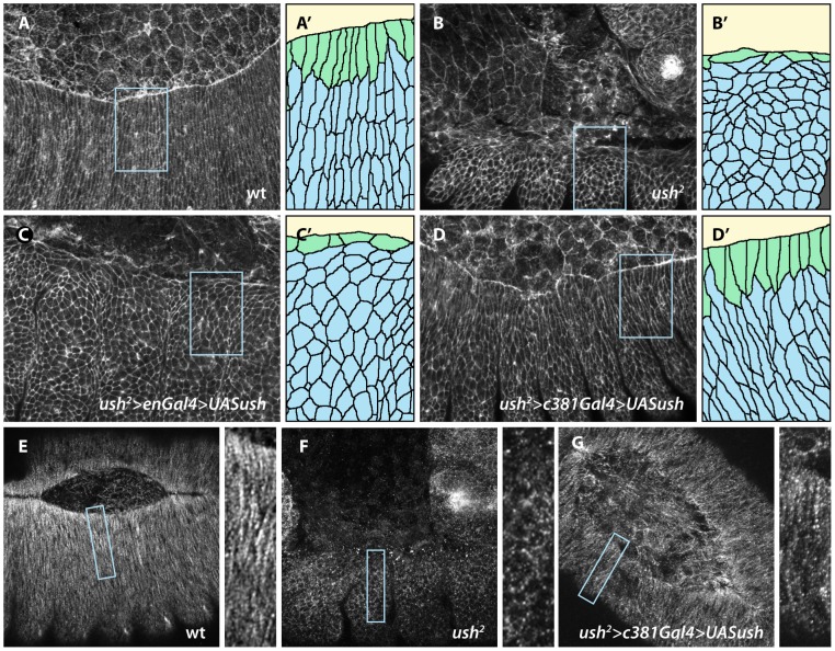
Fig. 3. An Ush-mediated signal from the AS induces elongation and D-V microtubule realignement of epidermal cells.
(A–D) Stage 14 embryos stained for F-actin. (E-G) Early stage 15 (E) and stage 14 (F,G) stained for α-tubulin. (A′–D′) Magnification of boxed region made by camera lucida tracing. (A) Wild type embryo. Epidermal cells are elongated in the D-V direction. (A′) DME cell are colored green, lateral epidermal cells blue. (B) ush2 embryos. Epidermal cells fail to elongate. (B′) DME and second row of epidermal cells are often stretched in the A-P direction while the bulk of epidermal cells tend to be isotropic. (C) Expression of Ush in epidermal stripes of ush2 mutants does not affect the shape of epidermal cells. (C′) Magnified region has been selected to cover a boundary between Ush-expressing and mutant epidermal cells and shows that the two first rows of epidermal cells still stretch in the A-P direction. (D) Expression of Ush in the amnioserosa of ush2 mutants rescues the elongation of epidermal cells. (D′) DME and lateral epidermal cells clearly elongate in the D-V direction. (E) Epidermal cell elongation is associated with formation of apical microtubule bundle that are oriented in the D-V direction, as shown here in wild type embryos. Right panel: magnification of the boxed region in (E). (F) Microtubules fail to form bundles and to orient in ush2 mutant embryos, Note their disorganized pattern in the corresponding magnification. (G) Restoring Ush in the amnioserosa of ush2 mutant embryos elicits correct bundling and D-V alignment of microtubules in the epidermis. All embryos are oriented with anterior to the left except embryo in G whose anterior end faces top left. Camera lucida drawings are 2× magnifications and the tubulin images are 2.5× magnifications of the corresponding boxed regions. All magnifications are as in Fig. 2 except for the insets in E, F and G, which are taken from the corresponding panels.

