Figure 2.

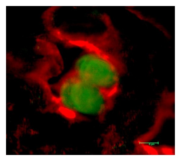
Skin sample from long-term WF-ACI VCA acceptor animals stained for CD4 (red) cell surface staining and FoxP3 (green) intracellular staining. The merged image shows CD4+/FoxP3+ cells.

Skin sample from long-term WF-ACI VCA acceptor animals stained for CD4 (red) cell surface staining and FoxP3 (green) intracellular staining. The merged image shows CD4+/FoxP3+ cells.