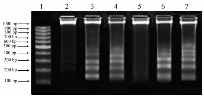
Figure 6.
DNA fragmentation induced by isolated PEs and PMA in both cell lines at CC50 concentration. The extracted DNA was run on 2% agarose gel and the image was documented using Bio-Rad Gel documentation system. Lane 1: 1 kb DNA ladder; Lane 2: un-treated Chang; Lane 3: Chang + PEs; Lane 4: Chang + PMA; Lane 5: un-treated Vero; Lane 6: Vero + PEs; Lane 7: Vero + PMA.

