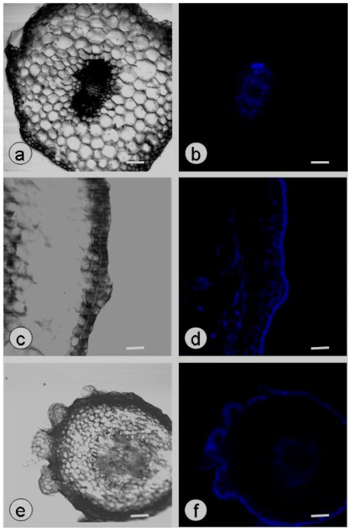
Figure 3.
Callose production in the hypocotyls of plasmolyzed zygotic embryos cultured on PGR-free MS. (a–f) Callose deposition was observed in the vascular tissues of untreated zygotic embryos (a,b), the epidermis and cortex of plasmolyzed zygotic embryos cultured for four weeks (c,d) and the globular somatic embryos and vascular tissues of plasmolyzed zygotic embryos cultured for six weeks (e,f). Confocal laser scanning microscope bright-field images (a,c,e) and aniline blue fluorescent staining of callose (blue; b,d,f); scale bar: 50 μm.

