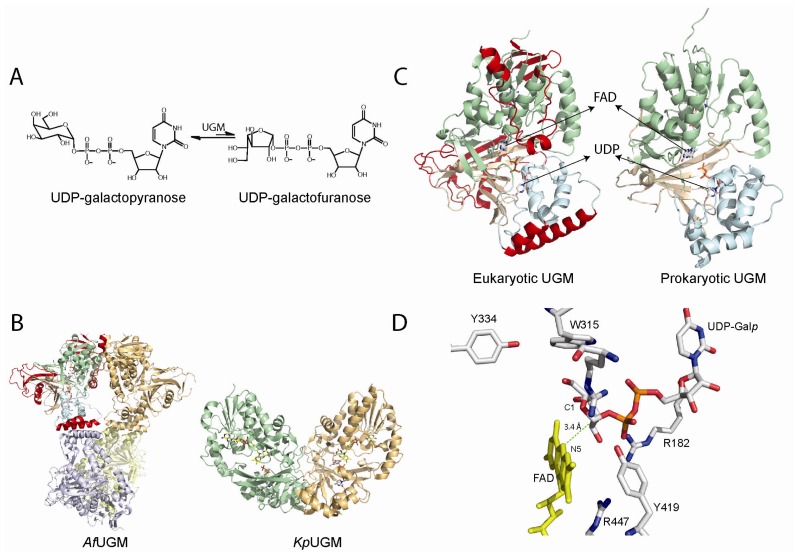
Figure 3.
(A) Reaction catalyzed by UDP-galactopyranose mutases (UGMs); (B) Cartoon representation of the tetrameric fungal (PDB code 3UTH) and dimeric bacterial (PDB code 3GF4) UGMs; (C) Monomers of UGMs showing the flavin adenine dinucleotide (FAD) binding domain (green), helical domain (blue), and beta sheet domain (gold). The additional secondary structures in the eukaryotic UGMs are highlighted in red; (D) Active site of eukaryotic UGMs showing residues predicted to interact with UDP-Galp. The interaction between the sugar C1 atom and the N5 of the FAD is depicted with a green dotted line.

