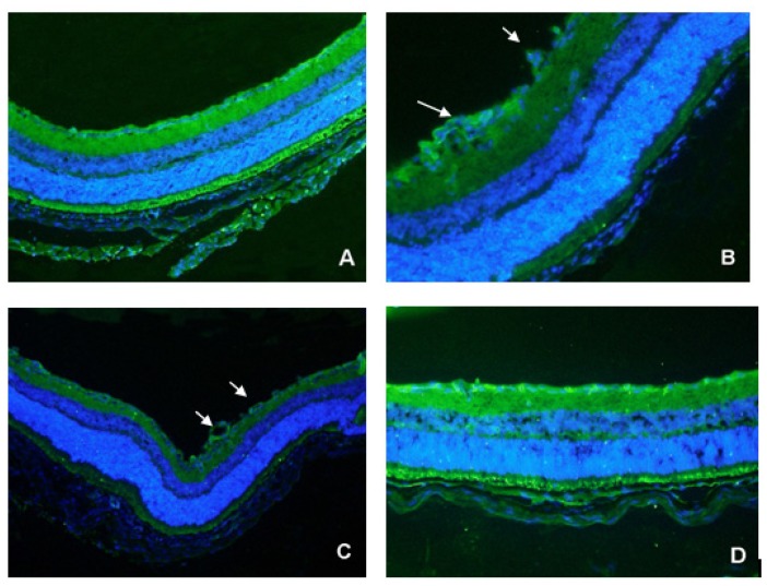
Figure 3.
Double immunohistochemical staining of frozen retinal sections of P17 C57BL/6 mice. Serial sections were treated with the polyclonal antibody, CD31. A green signal indicates positive staining for vessels, and blue signals indicate nuclei. (A) Normoxia condition; (B) Hyperoxia condition; (C) Injected with PBS before being exposed to 75% oxygen; (D) Injected with TeM before being exposed to 75% oxygen. Arrows: new blood vessels protruding into the vitreous cavity (B, C). Magnification: 100×.

