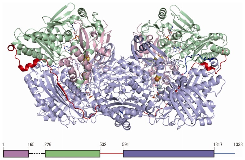
Figure 2.
Structure of human XOR. The structure illustrated is that of a human mutant dimeric XDH [69] (PDB: 2E1Q). The Fe/S, FAD, and molybdopterin domains are colored light pink, light green and light blue, respectively. The interdomain loop (residues 533–590) is colored red. C-terminal is colored blue. A schematic representation of the domain structure in relation to the primary sequence is shown at the bottom.

