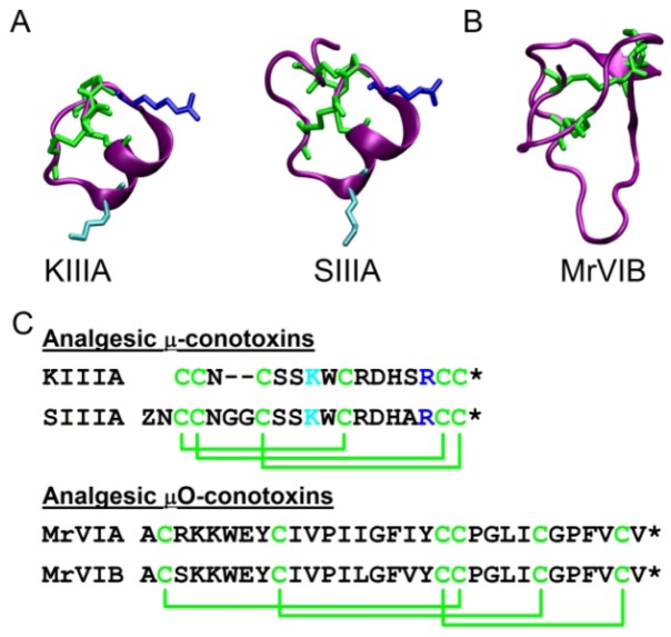
Figure 1.
Structures and sequences of analgesic μ- and μO-conotoxins. (A) Solution structures of analgesic μ-conotoxins, KIIIA and SIIIA. Disulfide links (green) with critical lysine (cyan, K7 (KIIIA) and K11 (SIIIA)) and arginine (blue, R14 (KIIIA) and R18 (SIIIA)) residues. (B) Solution structure of analgesic μO-conotoxin MrVIB. Disulfide links are green. (C) Amino acid sequences of analgesic μ-conotoxins, KIIIA and SIIIA, and μO-conotoxins, MrVIA and MrVIB. * C-terminal amidation.

