
Fig. 5.
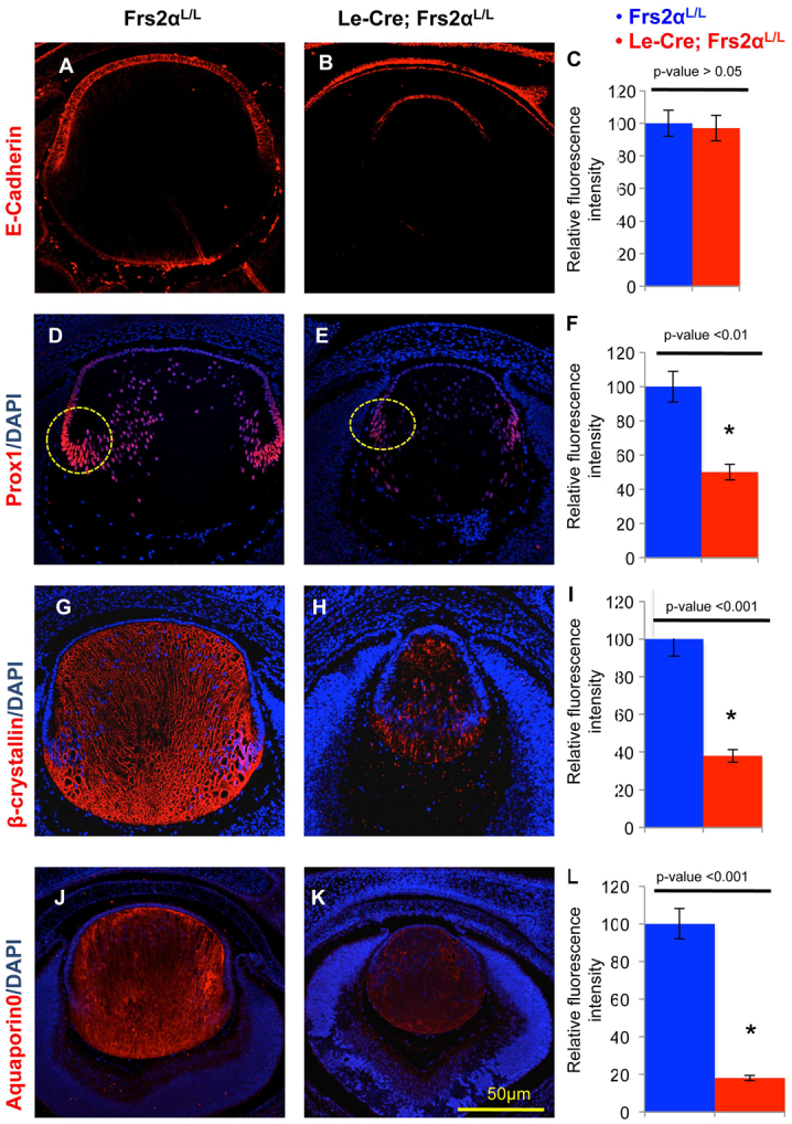
Frs2α-deficiency in the lens maintains lens identity but displays reduced levels of fiber differentiation-specific proteins. (A-L) Lenses from control (A,D,G,J) and Frs2α-deficient (B,E,H,K) E15.5 embryos were analyzed for E-cadherin (expressed in lens epithelial cells), Prox1, β-crystallin and aquaporin 0 (characteristic of lens fiber cells). Both the pattern (compare A with B) and expression level (C) of E-cadherin is similar in Frs2α-deficient and control lenses. Conversely, although the normal expression pattern of Prox1 (D,E), β-crystallin (G,H) and aquaporin 0 (J,K) were preserved, these proteins were significantly less abundant in the Frs2α-deficient lenses (F,I,L). Nuclear Prox1 fluorescence intensity was analyzed in the areas represented by the yellow circles in D and E. Error bars on the graphs represent s.e.m. Asterisks represent significant differences.