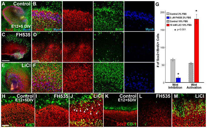
Fig. 3.
Modulation of Wnt/β-catenin signaling in the mitotic E12 prosensory domain affects proliferation and HC differentiation. (A-F) Low- (A,C,E) and high- (B,D,F) magnification lumenal surface views of E12 sensory epithelial explants maintained for 5 DIV with BrdU in control media (A,B), the Wnt/β-catenin inhibitor FH535 (C,D) or the Wnt/β-catenin activator LiCl (E,F). (B,D,F) Merged views are shown on the left and individual channels to the right. In controls, immunocytochemistry for Sox2 (red), BrdU (green) and Myo6 (blue) indicated that HCs develop normally and there is a moderate level of BrdU incorporation by Sox2-positive cells (A,B). Wnt inhibition results in a substantial reduction in HCs, BrdU incorporation and the size of the prosensory field (C,D), whereas Wnt/β-catenin activation substantially increases the number of HCs and BrdU-positive cells and the size of the prosensory field (E,F). (G) Quantification of Sox2+ BrdU+ cells within a 200×100 μm box positioned over the lateral HC domain at the basal-apical midpoint in control or Wnt-modulated explants after 4 DIV revealed a significant reduction or expansion in the number of cells that had undergone mitosis following treatment with Wnt inhibitor, or Wnt activator, respectively, compared with controls. Control samples maintained in low (2.5%) or high (10%) fetal bovine serum (FBS) appeared similar by immunocytochemistry, but were quantified independently; n=9 explants counted per condition; *P≤0.001 for both treatments. Error bars indicate ±s.e.m. (H-J) To identify ongoing proliferation, immunocytochemistry for Ki-67 (green) was performed on E12 control (H), FH535-treated (I) and LiCl-treated (J) explants maintained for 5 DIV. Almost no Ki-67-positive cells were observed in the Sox2-positive OC domain in control (H) and FH535-treated (I) cultures, whereas many were observed in LiCl-treated explants (J, arrowheads). (K-M) Expression of the cell cycle regulator cyclin D1 (CD-1, green) was observed just outside of the Sox2-positive (red) sensory domain in control explants (K), whereas CD-1 was almost completely absent from FH535-treated cultures (L). In LiCl-treated samples, CD-1 expression was increased and overlapped with the expanded Sox2-positive domain (M). Scale bars: 300 μm in A,C,E; 100 μm in B,D,F; 50 μm in H-M.

