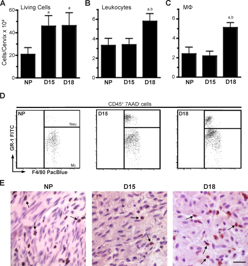
FIG. 3.
Mφ increase in the cervix before birth. The number of total living cells (7AAD–; A), leukocytes (CD45+; B), and Mφ (CD45+7AAD–F4/80+GR-1–; C) in the cervix as identified by flow cytometry and gated as described in Figure 2 is shown. Statistical significance (P < 0.05) for D18 (n = 5) versus groups of mice that were aNP (n = 6) or bD15 (n = 5) is also shown. D18 is pregnant mice on Day 18 postbreeding (n = 5). D) Representative plots of gated living Mφ and Neu in dispersed cervices from NP as well as D15 and D18 mice. E) Mφ stained dark brown by immunohistochemistry (arrows) and counterstained with hematoxylin in sections of cervix from NP and pregnant mice. Bar = 25 μm.

