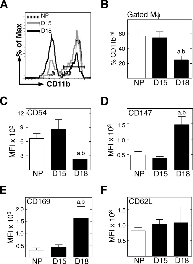
FIG. 4.
Mφ in the prepartum cervix increase expression of certain activation markers. Dispersed cells from perfused cervices were stained for flow cytometry, and a standardized gating protocol was used to identify F4/80+GR-1– Mφ. A) Representative histograms of CD11b expression by F4/80+GR-1− Mφ in the cervix of from mice that were NP, D15, or D18. B) Percentages of CD11b+hi Mφ/group (n = 3–5; Student t-test). C–F) Median fluorescence intensity of the various activation markers on F4/80+GR-1− Mφ/group (n = 4–6; Mann-Whitney test). For D18, statistical significance (P < 0.05) versus aNP or bD15 is shown.

