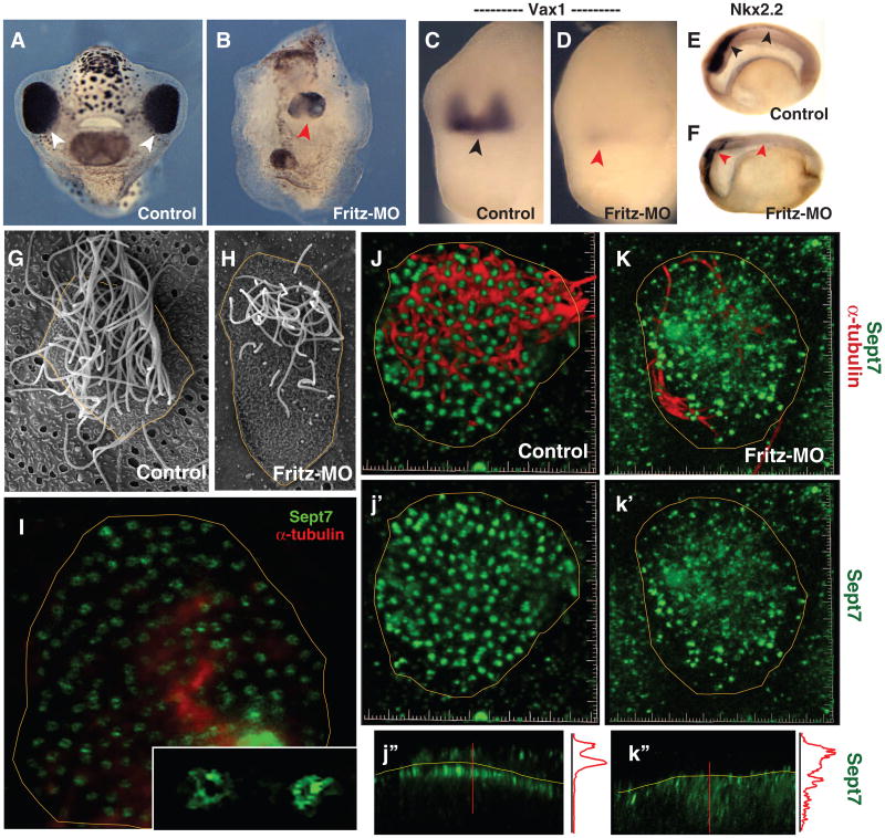
Fig. 3.
Fritz controls septin localization, ciliogenesis, and Hedgehog signaling.(A) Control tadpole (anterior view; arrowheads indicate eyes).(B) Sibling Fritz morphant (red arrowhead indicates single, medial eye). (C) Vax1 expression in control embryo.(D) Vax1 expression is lost in a Fritz morphant. (E) Sagittal view of Nkx2.2 expression in the spinal cord of a control embryo, black arrowheads. (F) Nkx2.2 expression is lost in Fritz morphant, red arrowheads. (G) A single multiciliated cell from the Xenopus epidermis. (H) Multiciliated cells in Fritz morphants display fewer and shorter cilia. (I) Sept7 in ring-like structures (inset) at the base of cilia in a confocal slice of a multiciliated cell. (J) Sept7 structures (green; j′) are highly ordered in a stack from a control multiciliated cell: cilia are visible in red. (K) Sept7 structures (green; k′) are disorganized in a Fritz morphant multiciliated cell: few cilia are visible in red. (j″) Z-projection reveals tight association of Sept7 with the apical surface (yellow line); intensity plot is shown at right (red line). (k″) Z-projection reveals loss of sept7 from the apical surface.

