
Figure 1.
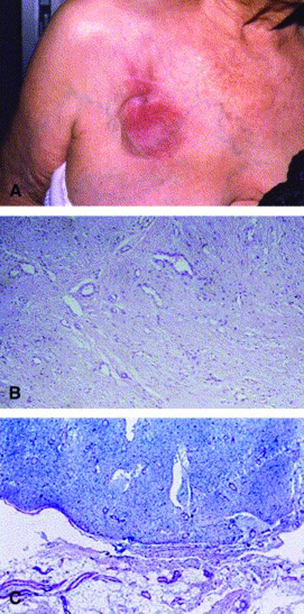
(A) Erythema-tous, slowly enlarging, ill-defined plaque on chest. Note prominent vessels on chest wall. (B) Histo-pathology showing diffuse reactive vascular hyperplasia (angiomatosis) in edematous, mucinous stroma. Note some prominent endothelial cells lining vessel walls. (Hematoxilin-eosin stain; original magnification x200.) (C) Abundant mucin deposition in dermal stroma associated with vascular hyperplasia and sparing subcutis. (Alcian blue, pH 2.5; original magnification x100.)
Reproduced with permission from the Journal of the American Academy of Dermatology.2