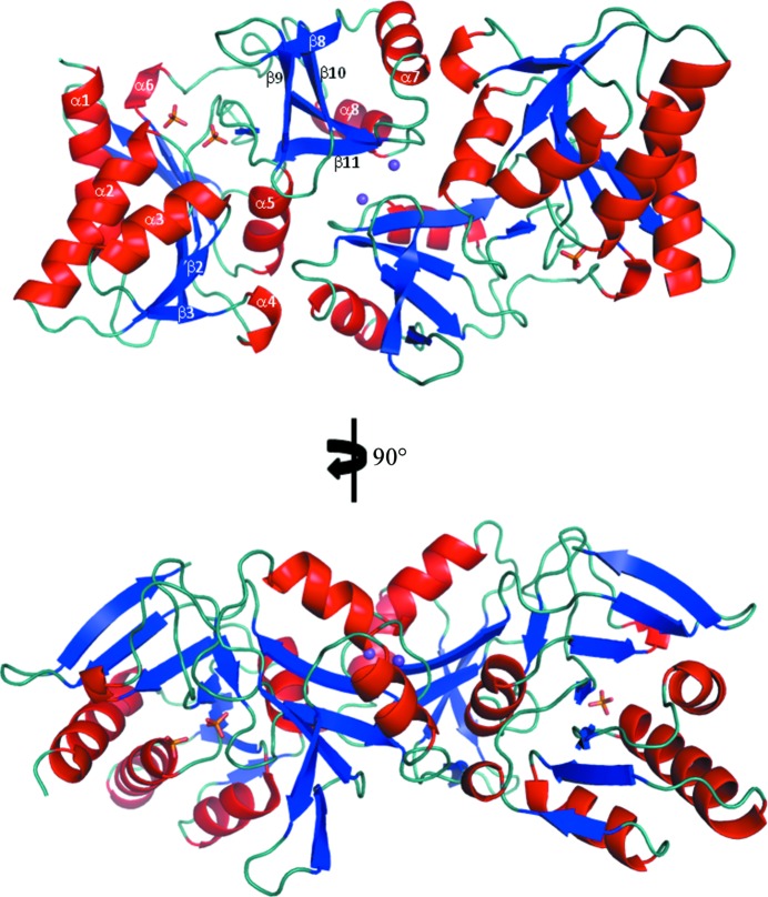
Figure 2.
The overall dimeric structure of RpiA from L. salivarius, with each chain coloured by secondary structure (helices in red, β-strands in blue and random coils in teal). The two K+ ions located at the dimer interface are shown as purple spheres and the three phosphate (PO4 3−) ions are shown in stick representation: two in the active site of chain A and one in the active site of chain B (yellow sticks).

