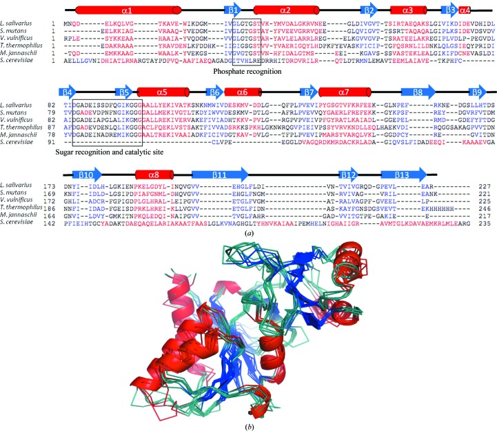
Figure 3.
(a) Structure-based sequence alignment of the L. salivarius structure with the published structures with PDB codes 3l7o, 3enq (Kim et al., 2009 ▶), 1uj4 (Hamada et al., 2003 ▶), 3ixq (Strange et al., 2009 ▶) and 1xtz (Graille et al., 2005 ▶). (b) Superposed structures of one protomer from the above members of the RpiA family coloured as in Fig. 2 ▶. The L. salivarius structure is shown in cartoon representation. Molecular-graphics figures were all prepared using PyMOL (Schrödinger LLC). The sequence alignment was prepared in PROMALS3D (Pei et al., 2008 ▶).

