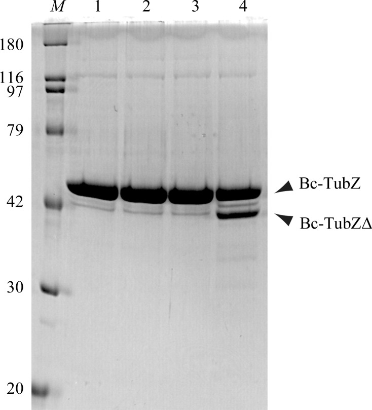
Figure 1.
Limited proteolysis of Bc-TubZ. Digested samples were resolved by 12% SDS–PAGE with Coomassie Blue staining. The major fragment is labelled with an arrow. The sizes of the molecular-mass marker (lane M) are indicated on the left in kDa. Lane 1, intact Bc-TubZ; lane 2, Bc-TubZ digested with trypsin; lane 3, Bc-TubZ digested with chymotrypsin; lane 4, Bc-TubZ digested with thermolysin.

