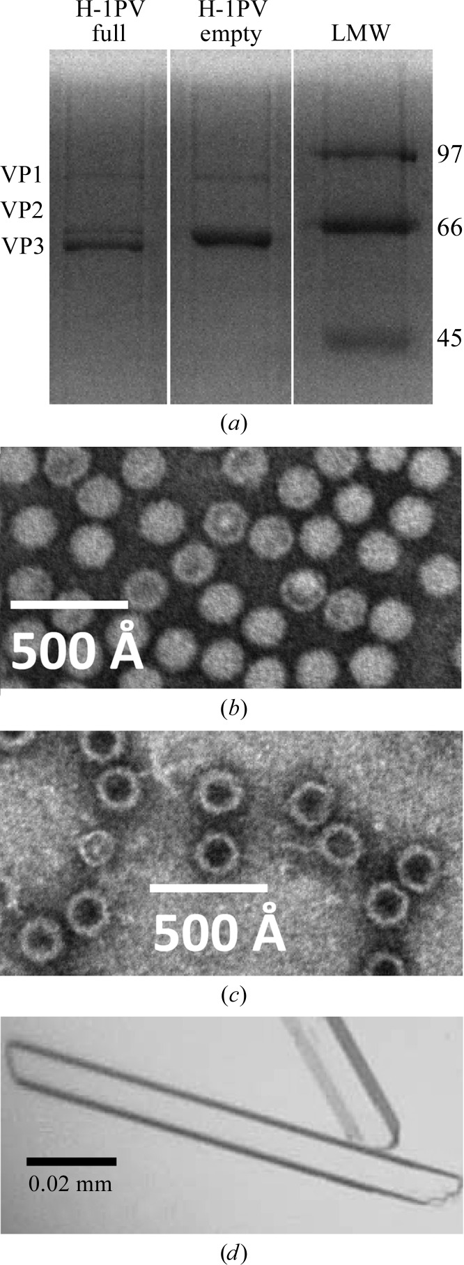
Figure 1.
Purification and crystallization of H-1PV full and empty capsids. (a) A 15% SDS–PAGE gel of the purified H-1PV capsids showing the relative ratios and positions of VP1, VP2 and VP3 (molecular weights of 81, 65 and 63 kDa, respectively). The positions of low-molecular-weight standards (labeled in kDa; Bio-Rad, Hercules, California, USA) are indicated on the right-hand side. (b, c) Negatively stained electron micrographs of H-1PV full capsids viewed at 100 000× magnification (b) and H-1PV empty capsids viewed at 60 000× magnification (c). (d) Optical photograph of H-1PV full capsid crystals in a rod-shaped habit.

