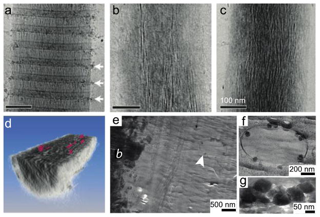
Figure 5.
Implementation of cryo-TEM to characterize transient biological organic-inorganic structures. To explore collagen mineralization, a model system with an amorphous mineral precursor was used. The collagen template was monitored over time by imaging the sample after (a) 24 hours, (b) 48 hours, and (c) 72 hours of mineralization. (d) A computer-generated three-dimensional representation from tomographic slices of a mineralized collagen bundle cut in cross section along the x–y plane to illustrate the plate-shaped apatite crystals (pink). To further investigate the role of amorphous calcium phosphate in developing mouse calvarial bone, (e) cryo-TEM of a vitrified section of tissue revealed the presence of intracellular amorphous calcium phosphate-containing vesicles (white arrowhead) in cells adjacent to the electron dense mineralized bone (labeled b). (f) A higher magnification of the vesicle in (e). (g) A freeze-dried cryo-section demonstrating the increased electron density from the mineral. Panels (a–d) reprinted with permission from [117]. Copyright 2010 Nature Publishing Group. Panels (e–g) reprinted with permission from [111]. Copyright 2011 Elsevier.

