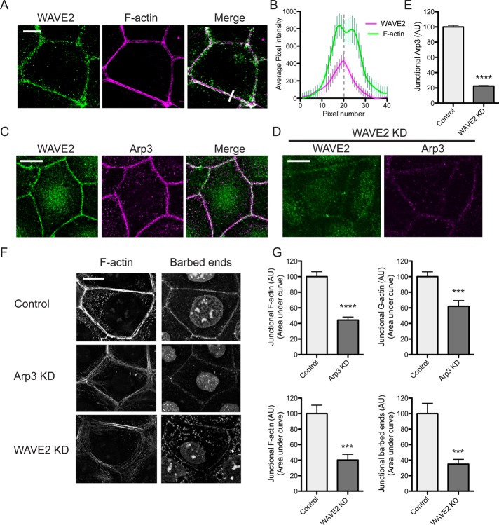
FIGURE 3:
WAVE2 regulates junctional actin assembly. (A, B) WAVE2 localized at the ZA between apical F-actin rings. WAVE2 and F-actin were visualized by confocal immunofluorescence imaging (A) and the fluorescence profiles measured by line scan analysis of 20 contacts (B). A representative line scan is marked by a white bar in the merged image in A. (C) Coaccumulation of WAVE2 and Arp3 at the ZA imaged by confocal immunofluorescence microscopy. (D, E) Junctional Arp3 is depleted in WAVE2 RNAi cells. Control and WAVE2 siRNA cells were immunostained for WAVE2 and Arp3. Costaining in WAVE2 KD cells is shown in D and junctional Arp3 quantitated by line scan analysis in control and WAVE2 KD cells in (E; n = 40 contacts from three independent experiments). (F, G) Junctional actin nucleation and F-actin are perturbed in Arp3 and WAVE2 RNAi cells. Barbed ends and junctional F-actin were labeled by incorporation of Alexa 594 G-actin and phalloidin staining, respectively, in control, Arp3 siRNA, and WAVE2 siRNA cells. Representative images are shown in F and quantitation in G. Data are n = 24 for control and n = 24 for Arp3 siRNA; n = 15 contacts for control and n = 13 contacts for WAVE2 siRNA from three independent experiments. ***p < 0.001; ****p < 0.0001; data are means ± SEM, normalized to control; AU, arbitrary units. Scale bars, 10 μm.

