FIGURE 5:
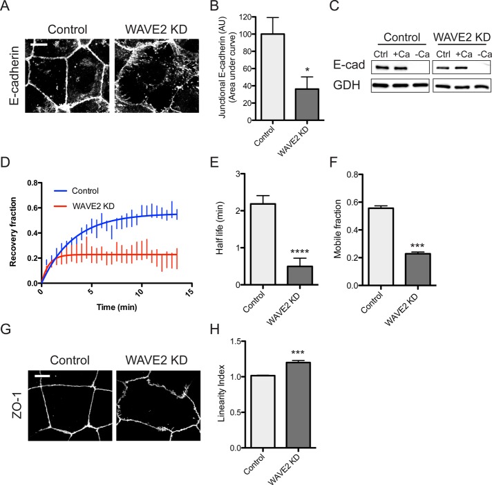
WAVE2 regulates E-cadherin organization and dynamics at the zonula adherens. (A, B) WAVE2 KD reduces apical concentration of E-cadherin into the ZA. Representative confocal immunofluorescence images of E-cadherin in control and WAVE2 KD cells (A) and quantitation by line scan analysis (B; n = 20 contacts from three independent experiments). (C) Total and surface E-cadherin (E-cad) levels in control and WAVE2 KD cells measured by sensitivity to digestion by extracellular trypsin in the presence or absence of Ca2+. Results are representative of three independent experiments. Ctrl, no trypsin added; +Ca, trypsin added in media containing 2 mM Ca 2+; –Ca, trypsin added in calcium-free media. GAPDH (GDH) was used as a loading control. (D–F) FRAP of junctional E-cadherin–GFP expressed in E-cadherin shRNA cells (n = 6). Half-life of recovery (E) and mobile fraction (F) were reduced in WAVE2 KD cells. (G) Junctional linearity was reduced in WAVE2 KD cells, as revealed by immunostaining for ZO-1 and quantitated (H; linearity index) as described in Materials and Methods. Data are means ± SEM, normalized to controls; n = 29 for control and n = 33 for WAVE2 KD from three independent experiments; AU, arbitrary units; ***p < 0.001, ****p < 0.0001. Scale bars, 10 μm.

