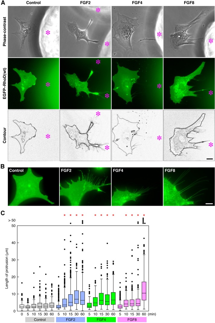
FIGURE 3:
RhoD(wt)-transfected cells extend thin protrusions by FGF stimulation. (A) Protrusion extension of RhoD(wt)-transfected cells toward FGF-coated beads. FGF2/4/8-coated heparin–acrylic beads were placed for ∼3 h in the culture of 10T1/2 cells transfected with EGFP–RhoD(wt) and then serum-starved. Asterisks indicate the position of FGF-coated beads. (B) Protrusion formation of RhoD(wt)-transfected cells stimulated with FGFs. EGFP–RhoD(wt)-transfected and serum-starved 10T1/2 cells were stimulated for > 60 min with FGF2/4/8. Scale bars: 10 μm. (C) The length of protrusions induced by FGF stimulation in RhoD(wt)-transfected cells. EGFP–RhoD(wt)-transfected and serum-starved 10T1/2 cells were stimulated with FGF2/4/8 for the time indicated. The length is shown as box-and-whisker plots with boxes and whiskers encompassing 75th/25th and 95th/5th percentiles, respectively. Dots out of the frame represent the number (but not the length) of >50 μm of protrusions. n > 110. *, p < 0.0001 by t test compared with the control.

