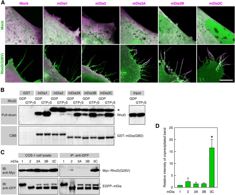
FIGURE 7:
mDia3C is localized to the RhoD-induced protrusions and binds to RhoD. (A) Distribution of mDia proteins in RhoD-transfected cells. The 10T1/2 cells were cotransfected with Cerulean–RhoD(G26V) (green) and EGFP–mDia proteins (magenta). Scale bar: 10 μm. (B) Binding of active RhoD to mDia1 and mDia3A/B/C in a pulldown assay. The binding of GDP- or GTPγS-loaded RhoD to GST–mDia(GBD) was analyzed by immunoblotting with the anti-RhoD pAb. *, an unidentified protein present in Sf21 cell lysates that was detected by the anti-RhoD pAb. (C) Specific binding of RhoD(G26V) to mDia3C in a coimmunoprecipitation assay. COS-1 cells were cotransfected with Myc–RhoD(G26V) and EGFP–mDia proteins, cell lysates were immunoprecipitated with anti-GFP pAb, and coprecipitated Myc–RhoD(G26V) was detected with the anti-Myc mAb. (D) Relative intensity of coprecipitated Myc–RhoD(G26V) band in the analysis of (C). The values are mean ± SD of three independent experiments. *, p < 0.05 by t test compared with mDia1.

