FIGURE 8:
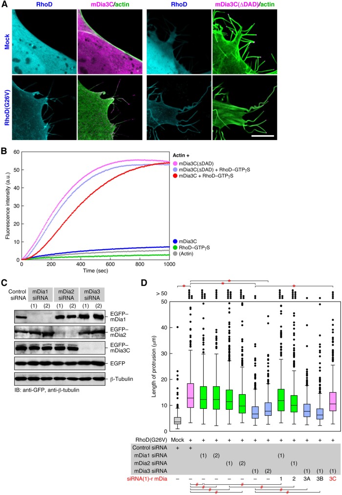
Activated mDia3C is essential for actin filament formation–mediated protrusion formation. (A) A constitutively active mDia3C(ΔDAD) forms protrusions independently of RhoD. The 10T1/2 cells were cotransfected with EGFP–mDia3C or EGFP–mDia3C(ΔDAD) (magenta) and mOrange2-Lifeact (green) together with or without Cerulean–RhoD(G26V) (cyan). Scale bar: 10 μm. (B) Facilitation of actin polymerization by mDia3C together with RhoD or solely by mDia3C(ΔDAD). Actin polymerization was analyzed by a fluorometric actin polymerization assay. a.u., arbitrary units. (C) Specific knockdown of mDia isoforms by respective mDia siRNAs. The 10T1/2 cells were first transfected with mDia Stealth siRNAs and 48 h later with EGFP–mDia1, mDia2, or mDia3C. The levels of proteins at 48 h after the second transfection were analyzed by immunoblotting with anti-GFP pAb. (D) Requirement of mDia3C but not mDia1, mDia2, or mDia3A/B for the RhoD-induced protrusion formation. The 10T1/2 cells were first transfected with mDia Stealth siRNAs and 48 h later with mCherry–RhoD(wt) and siRNA(1)-resistant EGFP–mDia1, mDia2, or mDia3A/B/C. The protrusion length was analyzed 48 h after the second transfection. n > 420. *, p < 0.0001, #, p > 0.01 (not significant) by t test.

