FIGURE 9:
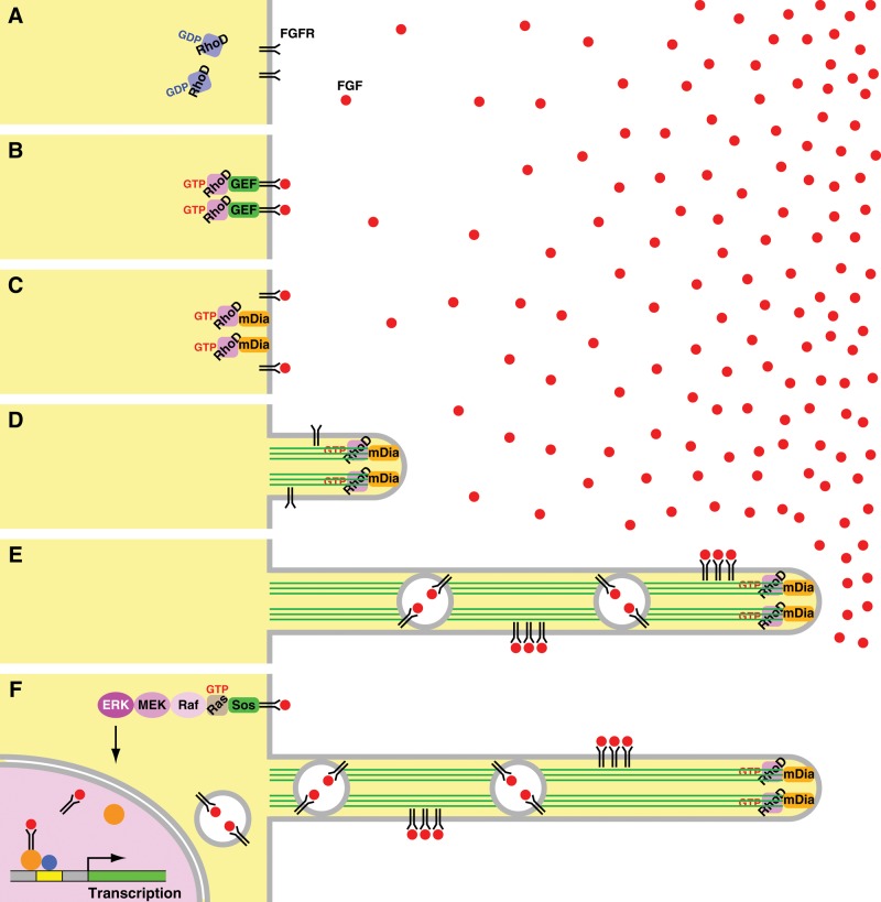
Postulated mechanisms of the FGF–RhoD–mDia3C-induced protrusion formation and the transport of FGFRs through the protrusions. (A) RhoD is inactive without FGF stimulation. (B) If FGF, even at a low concentration, binds to its receptors, a GEF for RhoD is activated by FGFR signaling. The GEF then activates RhoD. (C) Activated RhoD binds to activate mDia3C. (D) Activated mDia3C nucleates and elongates actin filaments to form protrusions. (E) Cells extend the protrusions toward high concentrations of FGF or the source of FGF. Higher concentrations of FGF can efficiently bind to FGFRs. FGF–FGFRs are transported through the protrusions either on the plasma membrane or on the early endosome-like vesicle membrane toward the cell body. (F) FGF–FGFRs transported to the cell body activates the canonical Ras–ERK, PI3K–Akt, or PLCγ signaling. FGF–FGFRs are also translocated to the nucleus through early endosomal compartments and participate in transcription of genes involved in cell growth, differentiation, or development.

