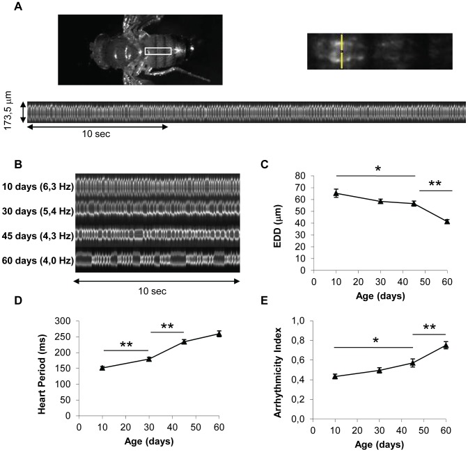
Figure 1. In vivo analysis of cardiac aging.
(A) Flies expressing a GFP protein targeted to mitochondria under control of the Heart-specific GeneSwitch protein (w/Y;UAS-mitoGFP/+; Hand-GS/+ male flies treated with RU486) were anaesthetized and fixed by their wings. Videos were acquired under a Stereomicroscope (1000 frames per movie, 32 frames/s). M-Mode was generated by horizontal alignment of rows extracted at the same position from each movie frame, with automated positioning of the acquisition zone (yellow segment). (B) Representative 10 second M-Modes of hearts at various ages. Cardiac frequency is indicated. (C,D,E) End Diastolic Diameter (EDD, µm), Heart Period (ms) and Arrhythmicity Index of 10-day-old (n = 26), 30-day-old (n = 34), 45-day-old (n = 34) and 60-day-old (n = 32) flies. All values are means (±SEM). Significant differences between successive ages are indicated: * p<5.10−2, ** p<5.10−3.

