
Figure 1. Structure of wild-type cardiac Troponin C.
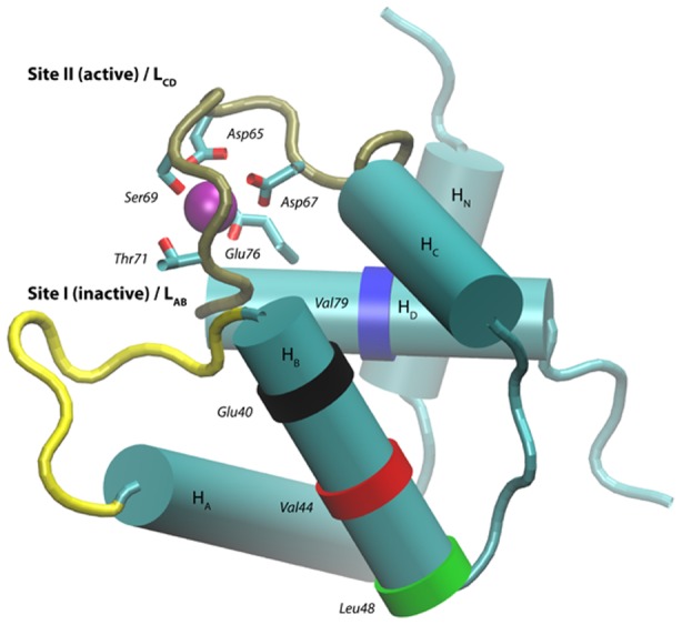
The physiologically inactive
 binding site I contains
  (yellow), while the active site II region contains
  (tan). The locations of the mutants in this study are marked as colored bands, including E40 (black) V44 (red), L48 (green) and V79 (blue).