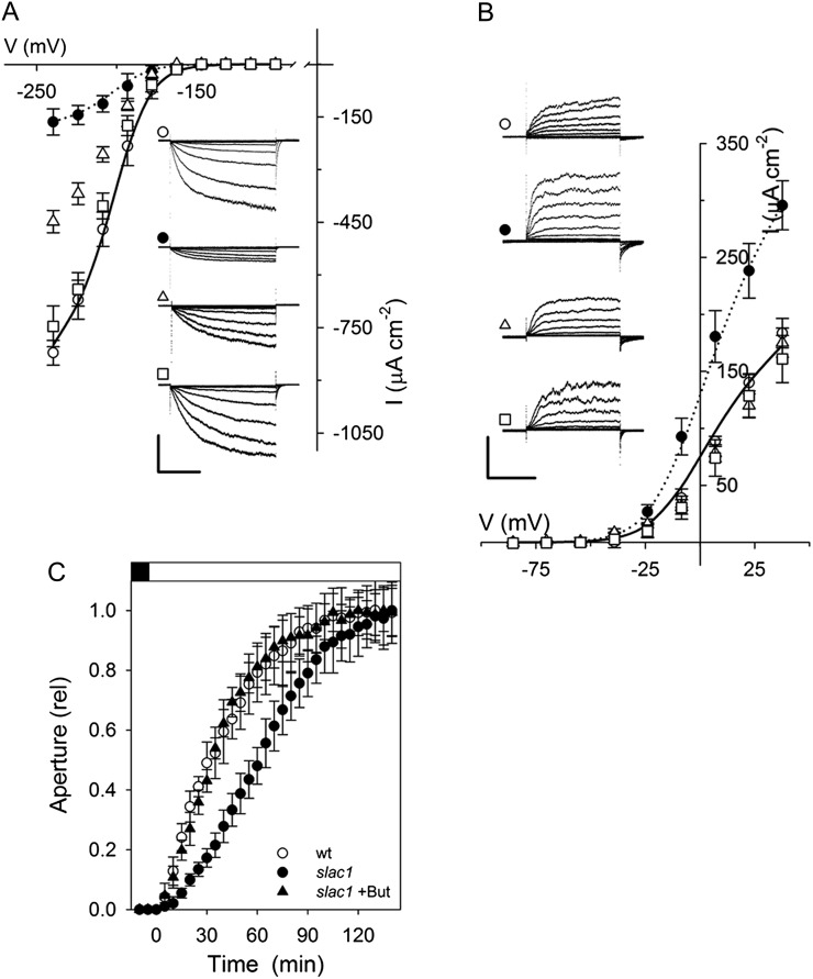
Figure 6.
Altered K+ channel currents are consequent on elevation of pHi and [Ca2+]i in the slac1-1 mutant. A, Mean ± se (n ≥ 8) of steady-state currents for IK,in under voltage clamp show recovery of wild-type characteristics in the slac1-1 mutant on suppressing [Ca2+]i and pHi elevations. Wild-type (open circles) and slac1-1 (closed circles) data are included from Figure 1 for reference. slac1-1 guard cells were loaded with 10 mm BAPTA to buffer [Ca2+]i (open triangles) and additionally exposed to 3 mm butyrate outside (open squares) to lower pHi (Fig. 5, A and B). The inset shows current traces recorded under voltage clamp and cross-referenced by symbol. Scales are as follows: vertical, 500 μA cm−2; horizontal, 2 s. B, Mean ± se (n ≥ 7) of steady-state currents for IK,out under voltage clamp show recovery of wild-type current characteristics in slac1-1 mutant guard cells on suppressing pHi elevation. Wild-type (open circles) and slac1-1 (closed circles) data are included from Figure 1 for reference. slac1-1 guard cells were exposed to 3 mm butyrate outside (open squares) to lower pHi (Fig. 5, A and B) and additionally when loaded with 10 mm BAPTA to buffer [Ca2+]i (open triangles). The inset shows current traces recorded under voltage clamp and cross referenced by symbol. Scales are as follows: vertical, 200 μA cm−2; horizontal, 2 s. C, Stomatal opening in epidermal peels on transition to 300 μmol m−2 s−1 light in the presence of 3 mm butyrate (But). Data are normalized to initial and final apertures after correcting for butyrate-induced aperture increase. Results for the wild type (wt) and the slac1-1 mutant are included from Figure 1 for comparison. Opening half-time was as follows: slac1-1 + butyrate, 23 ± 0.8 min. Apertures (initial/final in μm) are as follows: wild type, 2.7 ± 0.3/4.5 ± 0.3; slac1-1, 4.3 ± 0.3/5.1 ± 0.3; slac1-1 + butyrate, 4.5 ± 0.2/5.6 ± 0.2.

