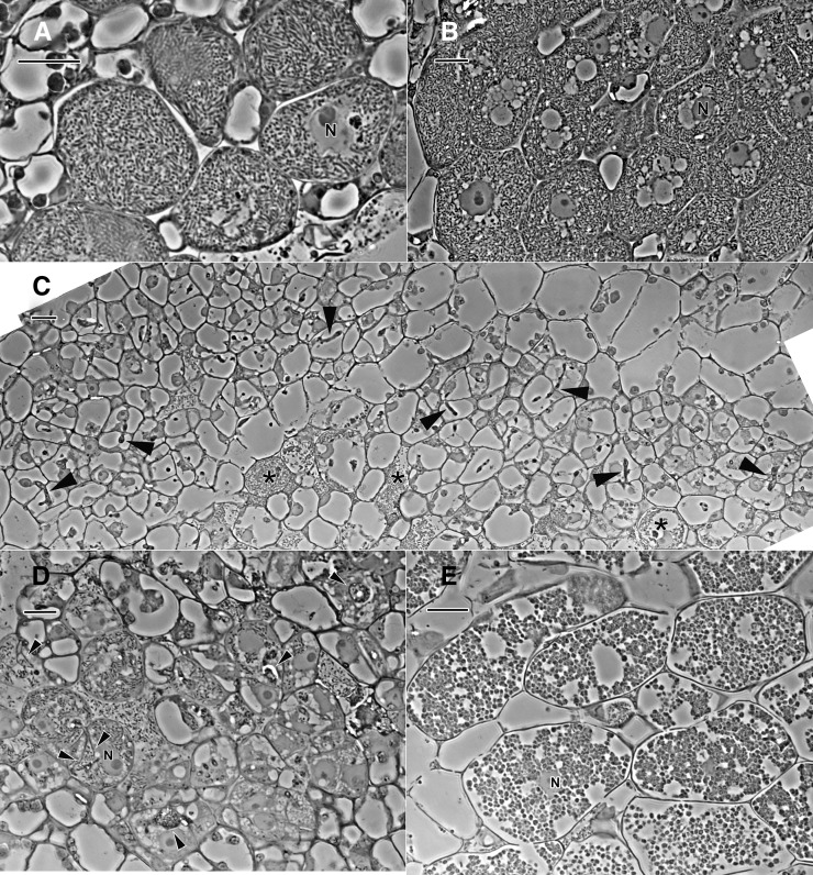
Figure 8.
Phase-contrast micrographs of 1-µm-thick resin sections of freeze-substituted nodules. A, GUS control. B, 14-3-3-silenced roots, medium-size nodule. C to E, 14-3-3-silenced roots, small-size nodule. N, Nucleus. The GUS control (A) and medium nodules (B) are similar in structure and are filled with typical symbiosomes. Small nodules contain cells that are arrested in development. In C, there are a few senescent host cells that contain rhizobia (asterisks), but most cells that have infection threads (arrowheads) are devoid of internalized rhizobia. There are more fully developed infected cells in the small nodule in D, but they are not as fully developed as those in medium nodules (B), and the infection threads (arrowheads) are distended with large numbers of rhizobia. Infected cells in the small nodule in E are filled with atypical symbiosomes that are spherical, and the host cytoplasm is degraded. Bars = 20 µm.

