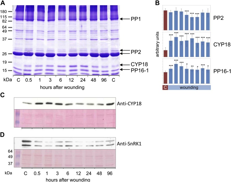
Figure 6.
PP2, CYP18, PP16-1, and SnRK1 are wound-regulated phloem proteins. A, Representative Coomassie blue-stained polyacrylamide gel displaying phloem proteins from untreated (two controls shown) or wounded pumpkin plants. Major phloem proteins PP1 and PP2, PP16-1, and CYP18 were labeled. CYP18 and PP16-1 were cut from the gel and identified by LC-MS/MS. B, Quantification of PP2 (major isoform), CYP18, and PP16-1 band intensities by ImageJ. Note the transient reduction in PP2 band intensity at 6 to 24 h after wounding, whereas CYP18 and PP16-1 were significantly induced from 0.5 to 96 h after wounding. Error bars indicate se (n = 9). Asterisks indicate statistically significant differences from the control (Student’s t test, *P < 0.05; **P < 0.01; ***P < 0.001). C, Western-blot analysis of phloem proteins with anti-CYP18 antibodies confirmed the wound induction of CYP18, although the kinetics is induction are somewhat variable. D, Western-blot analysis of phloem proteins with anti-SnRK1 antibodies. The western-blot signal corresponds to the expected protein mass of approximately 57 kD. The identity of the bottom band is unknown. The nitrocellulose membrane was stained with Ponceau red for confirmation of equal loading. C, Control. [See online article for color version of this figure.]

