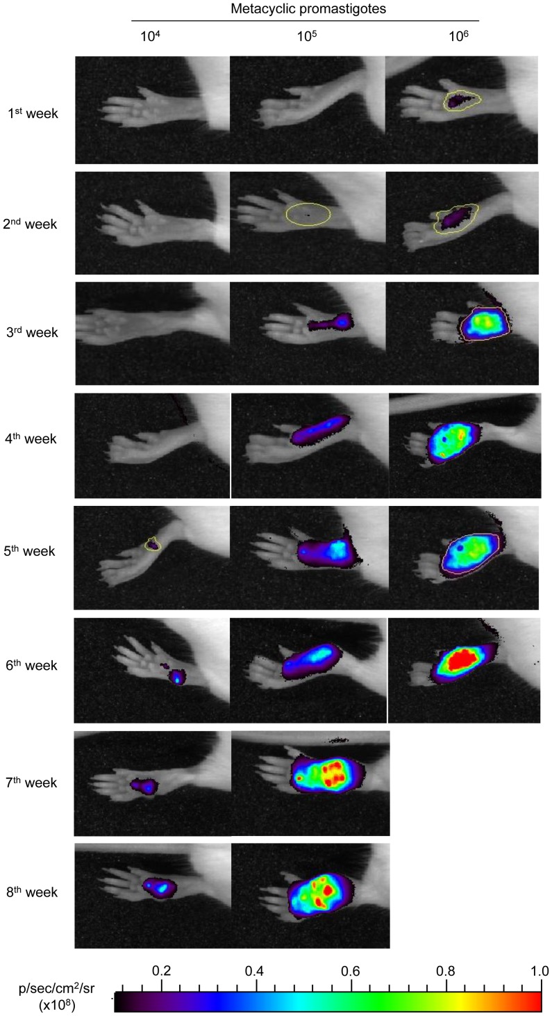
Figure 5. Progression of an experimental infection with mCherry+L. major in BALB/c mice.
Photographs of mouse footpads over the time after inoculation with 104; 105 and 106 mCherry+L. major metacyclic promastigotes. The images were taken weekly using an In Vivo Imaging System (IVIS 100; Xenogen) device. Six mice per dose were used in this experiment, and one representative mouse was chosen for all of the photographs. Examples of Regions of Interests (ROIs) used for quantification are marked in yellow.

