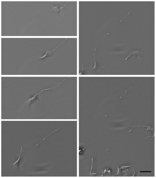
Figure 1. Time lapse acquisition of an isoquercitrin-stimulated NG108-15 cell.

The images, acquired every 56 minutes, show the migration path and neurite extension out of the cell body. Note that the neurite tail undergoes a small amount of translation, where the top of each image is at the same position. The video S1 is available in the Supplementary Information. Scale bar = 50 micron.
