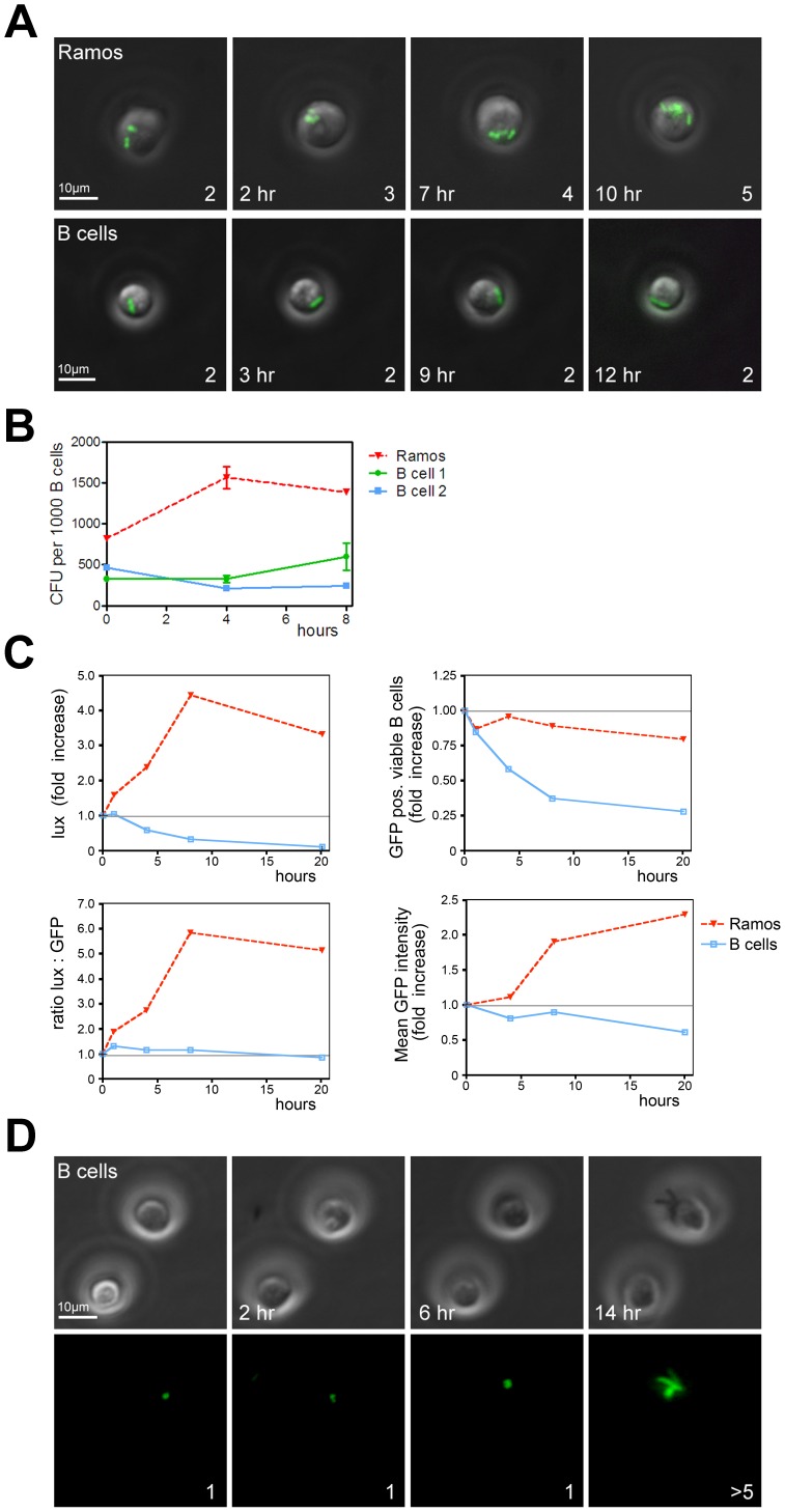
Figure 2. Primary human B cells form a survival niche for intracellular Salmonella.
(A) Widefield fluorescence microscopy of living Ramos and primary human B cells with phagocytosed anti-IgM coated GFP-Salmonella. Depicted are GFP signals projected on the transmission image. Scalebar = 10 µm. Number of bacteria in the visualized cell is given in the lower right corner. Lower left corner: time after Salmonella infection. Images are frames from Video S1 and S2. (B) Ramos and primary B cells were incubated with anti-IgM coated Salmonella, lysed and plated onto LB-agar plates at various time points after infection. Data are from triplicate experiments performed with Ramos and primary B cells from two individual healthy donors. Error bars indicate SEM. (C) Analysis of Ramos and primary human B cells incubated with living anti-IgM coated lux-expressing (top left panel) or GFP-expressing (top right panel) Salmonella. The ratio of lux over GFP shows the amount of light produced per GFP-Salmonella positive B cell (bottom left panel), indicating intracellular Salmonella viability. The mean fluorescence of the GFP positive population, set arbitrarily at 1 at the beginning of the experiment, shows that the GFP signal increases in Ramos B cells, whereas it decreased in primary human B cells (bottom right panel). A representative example of three independent experiments is shown. (D) B cells were infected with anti-BCR coated GFP-expressing Salmonella before exposure to Edelfosine to induce apoptosis. Cells were imaged over a 14 h period. Top panel: transmission image, bottom panel: GFP-signal. Scalebar = 10 µm. Images are frames from Video S3.

