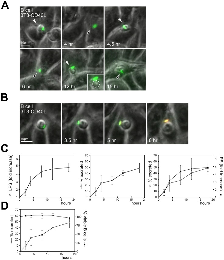
Figure 3. Salmonella is actively excreted by B cells.
(A) Primary B cells having phagocytosed anti-BCR coated GFP-Salmonella on a monolayer of 3T3-CD40L fibroblasts were imaged using widefield fluorescence microscopy. Depicted is the GFP signal projected on the transmission image with images taken every 30 min. Scalebar = 10 µm. Arrows indicate the B cell, white arrow: B cells moves op top of the monolayer, black arrow: B cells moves below the monolayer. Images are frames from Video S4. (B) Primary B cells having phagocytosed anti-BCR coated GFP-Salmonella on a monolayer of 3T3-CD40L fibroblasts were imaged using widefield fluorescence microscopy in the presence of TexasRed labeled anti-LPS mAbs. Depicted are GFP and Texas-Red signals projected on the transmission image. Scalebar = 10µm. Images are frames from Video S5. (C) Quantification of Salmonella secretion from B cells. Primary B cells were incubated with live uncoated GFP-Salmonella. Cells were stained with antibodies against LPS, fixed and analyzed using FACS. Left panel: increase in cell surface exposed LPS from bacteria exposed at the cell surface after initial uptake by B cells. Middle panel: percentage of B cells having excreted Salmonella as calculated from the percentage of B cells containing GFP-Salmonella followed in time. Right panel: left and middle panels are projected to illustrate that both processes show similar kinetics. Error bars represent SD from three independent experiments. (D) Primary B cells were incubated with live uncoated GFP-expressing Salmonella and followed for the time points indicated. The fraction of living B cells is plotted to demonstrate that loss of GFP-Salmonella positive B cells is not correlated with cell death.

