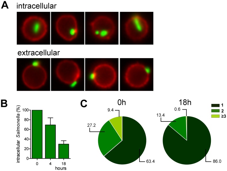
Figure 4. Quantification of the fate of the GFP-expressing Salmonella in infected B cells.
(A) B cells were infected with anti-BCR coated GFP-expressing Salmonella (green).The plasma membrane of the B cells (red) was stained using an anti-CD20 mAb to discriminate between intracellular and extracellular Salmonella. (B) The relative amount of intracellular Salmonella were measured immediately after infection (0 h), or 4 h and 18 h post-infection. Error bars represent SD from two independent experiments. (C) The number of Salmonella per B cell was measured immediately after infection and 18h post-infection. A representative experiment of two individual experiments is shown.

