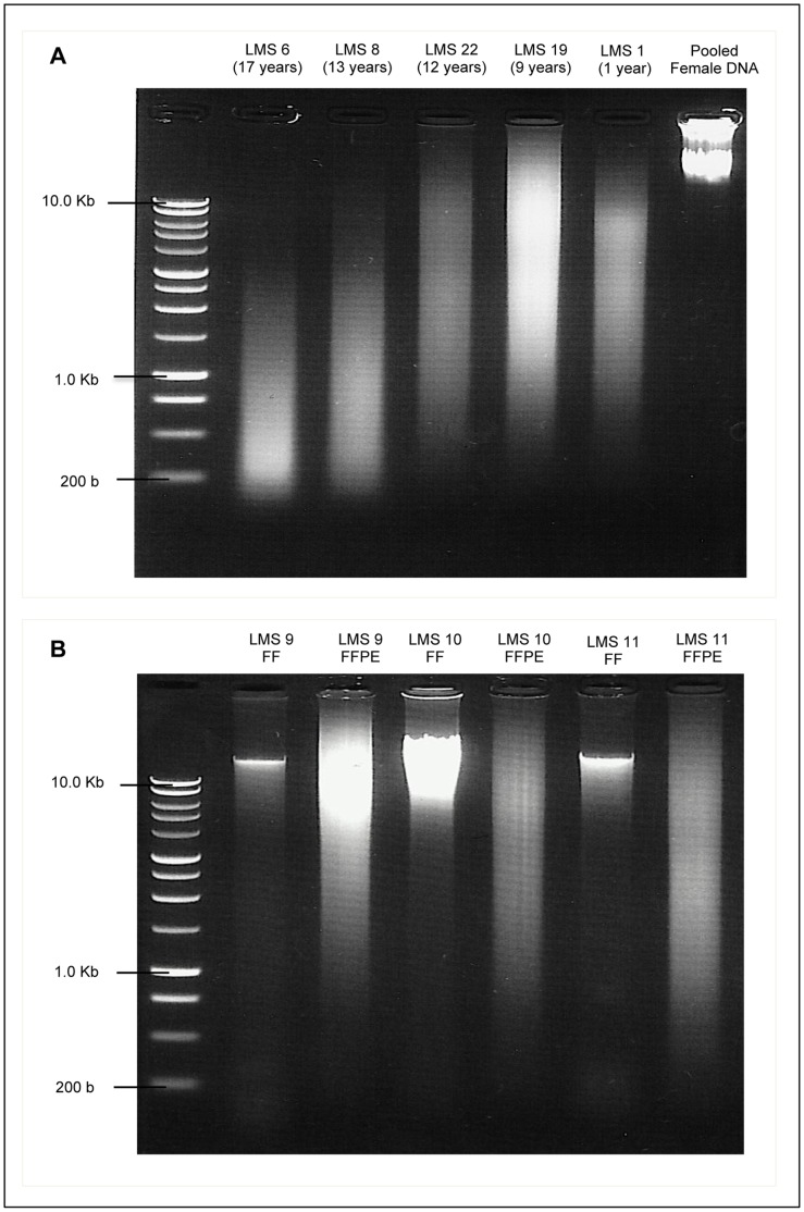
Figure 1. Agarose Gel images of DNA extracted from Leiomyosarcoma Tissue. A:
DNA extracted from FFPE leiomyosarcoma samples of different ages (shown in brackets) showing varied degrees of degradation, compared with commercial pooled female genomic DNA. B: Comparison of DNA extracted from paired FF and FFPE leiomyosarcoma samples (LMS 9, 10 and 11). FF samples show relatively distinct bands of high molecular weight, while corresponding FFPE samples show low molecular weight fragments in a wide range of sizes. All DNA samples are compared against a 1 Kb DNA ladder. DNA Electrophoresis was done on 1.0% agarose gels were pre-stained with Ethidium Bromide and examined under UV light.

