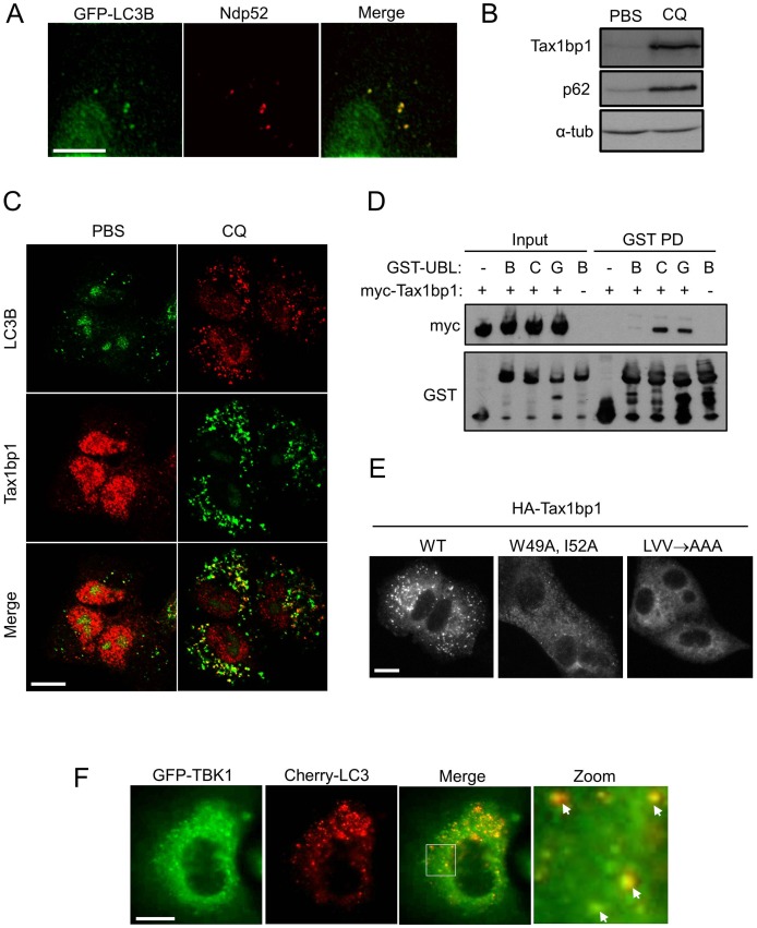
Figure 2. Basal autophagy targets TBK1-binding proteins Ndp52 and Tax1bp1. a).
A549 GFP-LC3B cells were stained for Ndp52 and imaged. b,c) A549 cells were treated with PBS or 5 µM chloroquine (CQ) overnight and b) cell extracts blotted for indicated proteins (α-tub = alpha tubulin) or c) cells co-stained for Tax1bp1 and LC3B, and imaged by confocal microscopy. d) 293FT cells were transfected with myc-TAX1BP1 expression vector, or empty vector control, and lysates subjected to pull-down with indicated fusion protein, as described in Materials and Methods (- = GST only, B = GST-LC3B, C = GST-LC3C, G = GST-GABARAP). e) A549 FLAG-HA-TAX1BP1 cell lines stably expressing the indicated variants of Tax1bp1 protein (see text) were treated with 5µM chloroquine overnight and stained for HA puncta. f) A549 GFP-TBK1 mCherry-LC3C cells were imaged (arrows indicate colocalisation). Scale bars = 25 µm in all panels.

