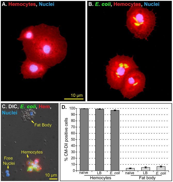
Figure 3. CM-DiI selectively stains hemocytes in vivo.
(A–B) Perfused hemocytes from mosquitoes injected with CM-DiI (red) and Hoechst 33342 (blue; nuclear stain). CM-DiI stains individual hemocytes (B) and hemocyte aggregates (A) from both naïve (A) and E. coli-infected (B) mosquitoes. (C) Bright field and fluorescence overlay of hemocytes, fat body and free nuclei collected by perfusion. Only hemocytes stain with CM-DiI. (D) Quantitative analysis of in vivo CM-DiI staining in perfused cells from naïve, injured (LB) and E. coli-infected mosquitoes. Greater than 97% of hemocytes stain with CM-DiI, fewer than 5% of fat body cells stain with CM-DiI, and free nuclei do not stain with CM-DiI. Columns, mean; bars, standard error of the mean.

