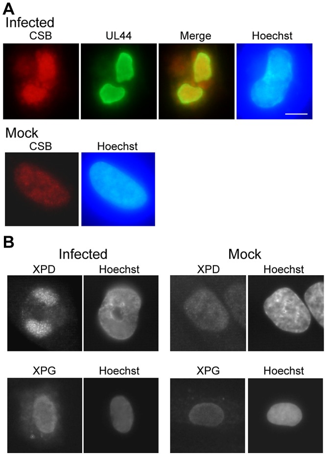
Figure 1. Certain NER proteins were tightly associated with HCMV RCs.

A) HFFs were mock- or virus-infected at an MOI of 5 for 48 h and harvested for IF analysis. Coverslips were treated using extract-first conditions (as described in Materials and Methods) and then stained simultaneously for CSB (red) and the viral processivity factor, UL44 (green). Overlay of the two images shows almost complete overlap of the signals (indicating tight association of CSB) in the infected cells. Staining of mock-infected cells shows nuclear localization of CSB. DNA was counterstained with Hoechst (blue). Scalebar on all images = 5 µm. B) Cells were stained for XPD (using “fix first conditions) in the top panels or for XPG (using “extract-first” conditions) in the bottom panels. Mock-infected cells again show nuclear localization of these antigens.
