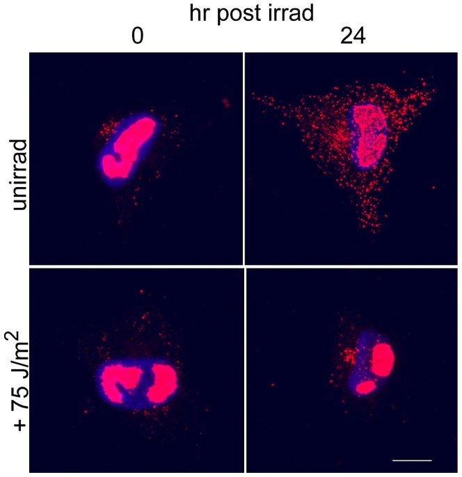
Figure 6. BrdU pulse and chase of viral DNA revealed no appreciable migration of pulsed viral DNA out of RCs during 24 h chase.

HFFs were infected on coverslips. One h prior to irradiation at 48 hpi, infected cells were pulse-labeled with BrdU. One half of the coverslips were then irradiated with 75 J/m2. The second half was not irradiated. Timepoints were taken at 0 and 24 h post irradiation (or control treatment). Coverslips were fixed and stained for BrdU incorporation and imaged using confocal microscopy. Projection images of the entire stacks are shown, with Hoechst staining of the nuclei in blue.
