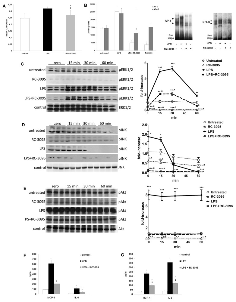
Figure 1.
RC-3095 decreased TLR4 mRNA expression, signaling pathways and cytokine/chemokine expression in RAW 264.7 cells exposed to LPS. Cell cultures were exposed to LPS (100 ng/mL) and, 4 h later, RC-3095 (10 ng/mL) was added for 2 h. Several times after this period, cells and/or media samples were collected for analysis in RC-3095–free media. (A) RT-PCR, using specific primers to TLR-4, demonstrated that 2-h RC-3095 treatment of LPS-activated RAW 264.7 cells (collected 30 min after RC-3095 treatment) reduced TLR-4 mRNA levels (expressed as the ratio of signal intensity to that of coamplified GAPDH: TLR-4 mRNA/GPDH) (*p < 0.05 versus control and $p < 0.05 versus LPS without RC-3095). (B) RAW 264.7 macrophages were stimulated with LPS; and 30 min after RC-3095 was added, LPS increased NF-κB and AP-1 DNA-binding activity measured by EMSA; and this was attenuated by RC-3095 treatment (*p < 0.05 versus untreated, $p < 0.05 versus LPS). (C–E) Western blot experiments showed that RC-3095 treatment also resulted in a sustained (0–60 min) reduction of pERK1/2, phosphorylated JNK and phosphorylated Akt levels (*p < 0.05, **p < 0.001 and ***p < 0.0001 versus untreated; ap < 0.05 versus LPS). (F) ELISA showed that RC-3095 attenuated MCP-1 and IL-6 increases induced by LPS stimulation in RAW 264.7 cells (G) and in peritoneal macrophages 30 min after the end of treatment (*p < 0.05 versus control and $p < 0.05 versus LPS).

