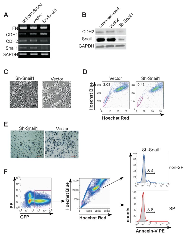
Figure 6.
Downregulation of Snail1 decreased ovarian cancer stemlike cell percentage and inhibited their invasive capacity. Untransduced cells, and cells transduced with control vector or shSnail1 (all from HO-8910PM cells) were analyzed for expression of CDH1, CDH2, FN and Snail1 by RT-PCR (A), Western blotting assay (B) and were detected under microscope (C). Scale bar = 50 μm. (D) SP cells were determined by FACS analysis in HO-8910PM cells transduced with control vector or shSnail1 lentivirus. (E) HO-8910PM cells transduced with control vector or shSnail1 were suspended in serum-free media and plated onto top chamber of Matrigel-covered micropore transwell. Cells migrated into membrane were stained with Hematoxylin and counted under microscope. Scale bar = 100 μm. (F) Experimental scheme for FACS analysis of apoptosis in lentiviral transduced shSnail1 cells. GFP-positive (transduced) cells were analyzed (left) and gated as SP cells and non-SP cells (middle). Percentage of apoptotic cells in GFP-positive SP cells (red) or GFP-positive non-SP cells (blue) were determined by PE-conjugated annexin-V staining (right). The histogram curves were overlaid by the SP cells or non-SP cells without annexin-V staining (tint).

