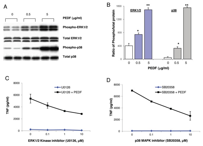
Figure 2.
PEDF induces inflammatory signaling pathways in murine macrophages. (A, B) RAW were cultured with different concentrations of recombinant PEDF. Total cell lysates were resolved by sodium dodecyl sulfate–polyacrylamide gel electrophoresis (SDS-PAGE) and immunoblotted with antibodies to phosphorylated and total ERK1/2 and p38 MAPK. Data are mean ± SE. *p < 0.05 and **p < 0.005 versus medium-alone control. (C, D) RAW cells were cultured with recombinant PEDF for 2.5 h in the presence or absence of ERK1/2 inhibitor (U0126) or p38 MAPK inhibitor (SB20358) at the indicated concentrations. TNF levels were measured in the supernatant by ELISA. Data are mean ± SE.

