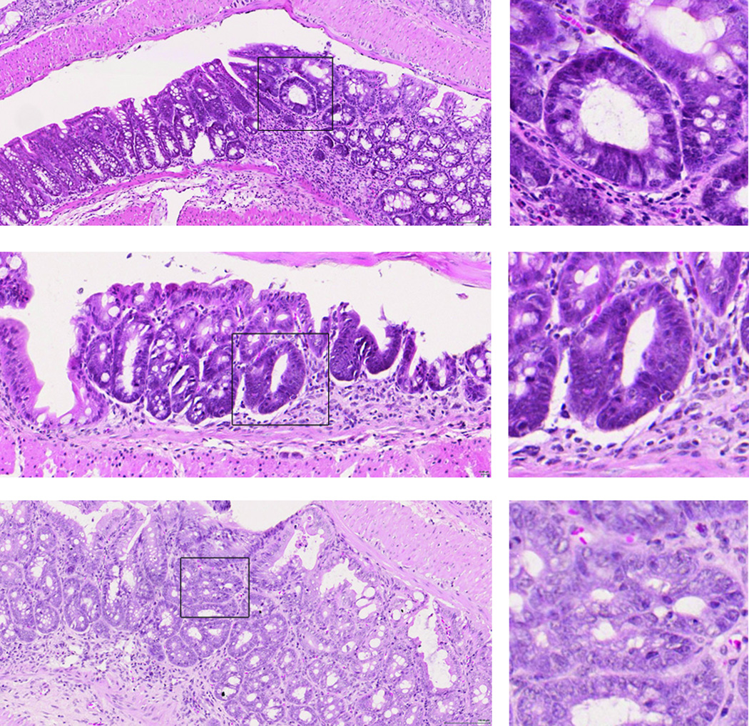
Fig 6. Dysplastic lesions are present in the colons of MMP10−/− mice after two rounds of DSS treatment.
Shown are examples of hematoxylin and eosin-stained lesions from 3 different MMP10−/− mice. Boxed regions showing examples of dysplasic lesions are shown in higher magnification on the right. Scale bar = 100 µm.

