
Figure 13.
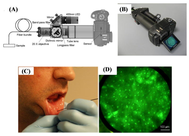
Schematic diagram (A) and picture (B) of the portable fiber-optic fluorescence imaging platform that uses a digital single-lens reflex (DSLR) camera introduced by Shin et. al.124 (C) In vivo imaging of healthy human oral mucosa. (D) An image of human mucosa that is labeled by proflavine were acquired by the DSLR based micro-endoscope shown in (A) and (B). Reprinted from Ref. 124 with permission from PLoS One.